Pelvis Bone Diagram. The pelvic cavity primarily contains the reproductive organs urinary bladder distal ureters proximal urethra terminal sigmoid colon rectum and anal canal. They help the lungs move and assist in the process of breathing.
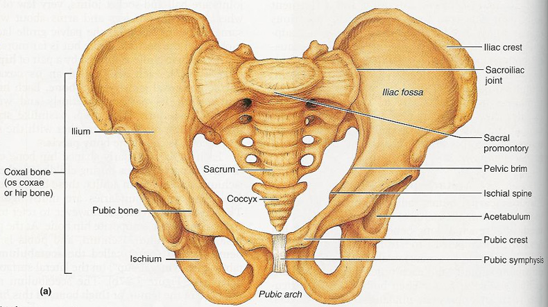
The tensor fasciae latae muscle attaches to the lateral aspect of the superior anterior iliac spine and also. Female pelvis bones. The bones of the pelvis are the hip bones sacrum and coccyx.
The pubic symphysis and the sacroiliac joint and reinforced by pelvic muscles.
Bone growth diagrams show the progression of development of the bone over a period of time. The tensor fasciae latae muscle attaches to the lateral aspect of the superior anterior iliac spine and also. This visually displays where a bone accepts blood vessels or where cartilage develops. We would like to show you a description here but the site wont allow us.
