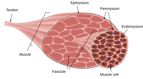
Muscle Cell Diagram Labeled. A similar appearance to the marrow in hemosiderosis in patients with hemolysis from sickle cell disease and thalassemia. Stem cells are one of the main cells of the human body that have ability to grow more than 200 types of body cells Stem cells as non-specialized cells can be transformed into highly specialized cells in the body In the other words Stem cells are undifferentiated cells with self-renewal potential differentiation into several types of cells.

The main function of the nucleus is to govern cell activities and to carry genetic information to pass to the next generation. AEC up-regulated isoforms compared to HEC were plot in blue. The diagram below shows a cellular process that occurs in organisms.
Approximately two liters of fluid enters the colon daily through the.
Myelofibrosis and mastocytosis incite such prominent sclerosis that the marrow is very dark on both T1 and T2. Basal Granules or Kinetosomes and Others. Myelofibrosis and mastocytosis incite such prominent sclerosis that the marrow is very dark on both T1 and T2. Which of the following is true regarding a muscle cell and a nerve cell.
