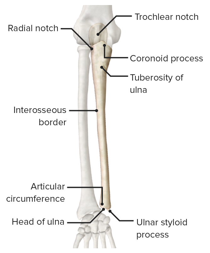
Medial Bone Of Forearm. The muscles of this chapter are involved with motions of the forearm radius and ulna at the radioulnar joints the hand at the wrist radiocarpal joint and the fingers at the metacarpophalangeal MCP andor the proximal interphalangeal PIP and distal interphalangeal DIP joints. This quiz is unlabeled so it will test your knowledge on how to identify these structural locations trochlea coronoid fossa deltoid tuberosity medial epicondyle lateral supracondylar ridge radial groove.

This nerve is directly connected to the little finger and the adjacent half of the ring finger innervating the palmar aspect of these. Distal Radial Glide on Ulna bone edit edit source Patient positioned supine with the arm at the side forearm in neutral. The outer surface of the muscle lies against the inner surface of mandible from which it is separated by the lateral pterygoid muscle sphenomandibular ligament maxillary artery mandibular nerve and its lingual and.
Distal Radial Glide on Ulna bone edit edit source Patient positioned supine with the arm at the side forearm in neutral.
Flexor-pronator tendon degeneration occurs with repetitive forced wrist extension and forearm supination during activities involving wrist flexion and forearm pronation. In human anatomy the ulnar nerve is a nerve that runs near the ulna bone. Thereby tendon degeneration appears instead of repair. The medial epicondyle of the humerus is an epicondyle of the humerus bone of the upper arm in humans.
